Improvement of the dosimetric properties of chemical-vapor-deposited
diamond films by neutron irradiation
| Mara Bruzzi,a) David Menichelli, and Silvia Pini Dipartimento di Energetica, Via S. Marta, 3, I-50139, Florence, Italy and INFM, Via G. Sansone, 1, I-50018 Sesto Fiorentino, Italy |
| Marta Bucciolini Dipartimento di Fisiopatologia Clinica, Firenze, Italy and INFN, Via G. Sansone, 1, I-50018 Sesto Fiorentino, Italy |
| József Molnár and András Fenyvesi Institute of Nuclear Research of the Hungarian Academy of Sciences (ATOMKI), Bem tér 18/c., P.O. Box 51, H-4001 Debrecen, Hungary |
(Received 13 March 2002; accepted for publication 6 May 2002) |
The performance of chemical-vapor-deposited (CVD). diamond films as on-line dosimeters has been substantially improved after irradiation with fast neutrons up to a fluence of 5 ×10¹⁴ n/cm2. This is correlated to a decrease of more than one order of magnitude in the concentration of deep levels with activation energy in the range 0.9–1.4 eV, as observed by thermally stimulated current and photoinduced current transient spectroscopy. As a consequence, a fast and reproducible dynamic response is observed during irradiation with a 6 MV photon beam from linear accelerator and with a Co60 source. A quasilinear dependence of the current on the dose rate is obtained in the range of interest for clinical applications (0.1–10 Gy/min). The resulting sensitivity is definitely higher than that of standard ionization chambers, and compares favorably with those of standard silicon dosimeters and of best-quality natural and CVD diamond devices. © 2002 American Institute of Physics. [DOI: 10.1063/1.1491014] |
Many outstanding properties of diamond, in particular soft-tissue equivalence, chemical inertness, nontoxicity and radiation hardness, make this material extremely appealing for clinical dosimetry.1–3 On-line dosimeters made with natural diamond are already commercially available but extremely expensive and rare due to the difficulty in selecting stones with the proper dosimetric characteristics. Chemicalvapor-deposited (CVD) diamond films have been recently proposed in their stead, due to the potential low cost of this material. One crucial problem still unsolved is the CVD dia mond doping recipe required to optimize the performance of such devices. It is well known that a quasi-uniform distribution of deep levels is needed to ensure the linear response of the current signal as a function of the dose rate;4 unfortunately, a high concentration of deep levels might also lead to a worsening of the device transport properties. The bulk of undoped CVD diamond films is characterized by a complexly structured distribution of traps giving rise to many active energy levels. Luminescence spectra are dominated by the broad A band,5 placed near 3 eV, now related to defects (possibly sp2-like structures) at dislocations and grain boundaries.6 However, electrical properties at room temperature are mainly influenced by shallower levels, below 1.5 eV, due to incorporated impurities or correlated to extended defects at grain boundaries.7,8 These traps can be effective in reducing the carrier lifetimes and, consequently, in worsening the sensitivity, the dynamic response, and the reproducibility of the device during exposure to the radiotherapy beams.9
In this work, we demonstrate that it is possible to
significantly improve the dosimetric performance of CVD diamond
films by opportunely tailoring the deep level distribution
through a preirradiation stage with fast neutrons at high
fluence levels. A commercial CVD diamond film with
1 × 1cm2 area and 600 μm thickness, equipped with Cr/Au
ohmic contacts (5 mm diameter) has been irradiated at
ATOMKI, Debrecen, Hungary, using fast neutrons produced
by 16 MeV protons in a 3 mm thick beryllium target and up
to a fluence of 5 ×10¹⁴ n/cm2. The neutron energy spectrum
is monotonically decreasing in the range from 0.1 MeV to
about 15 MeV; for higher energies a sharp cutoff occurs.10
Deep levels before and after neutron irradiation (NI) have
been investigated by means of thermally stimulated currents
(TSC) and photoinduced current transient spectroscopy
(PICTS) using a xenon lamp which generates pulses of 0.1 J
2 µs duration, covering the wavelength range 200–1000 nm,
with a peak at 300 nm. The TSC spectra, measured
before and after NI, exhibit a broad line peaked at ∼520 K,
as shown in Fig. 1. A detailed investigation revealed that this
dominant feature is due to the emission from different deep
levels.11 At least four components are needed to fit the
experimental results. The best fit is obtained using four deep
levels (Nos. 1–4) characterized by activation energies
in the range 0.9–1.4 eV, small capture cross
sections (10-17–10-19 cm2) and total concentration Nt
≈19 cm-3. Illumination with the Xe lamp does not ensure
a complete trap priming; thus, in order to completely saturate
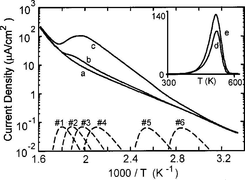
FIG. 1. TSC measurements performed with heating rate β=0.15 K/s and Vbias=5100 V. The background current (a) is measured without excitation. The other curves in the main plot correspond to measurements perfomed after a 30 min excitation using the Xe lamp, before (c) and after (b) NI. The contributions of fit components Nos. 1–6, calculated with arbitrary amplitudes, are shown in the bottom (dashed lines) for comparison. The spectra in the inset have been measured after a priming with a Co60 γ-source, up to a dose of 8 Gy; the signal measured after NI (d) is multiplied by ten for better comparison with the spectrum measured before NI (e).
the dominant levels, the sample has been irradiated with the Co60 source up to a dose of 8 Gy. The resulting TSC spectra are plotted in the inset of Fig. 1, showing that the spectral amplitude is reduced by a factor 20–30 after NI. Some defects activate near room temperature, generating a spectral contribution in the range 300–400 K. The total concentration of these traps before NI was Nt≈1016 cm-3, but after NI they are no longer observable by TSC. They have been also studied by PICTS, which offers higher sensitivity and resolution than TSC: Results are shown in Fig. 2. The PICTS spectra is calculated as S(T)=J(t2,T)-J(t1,T); J(t,T) denotes the current density transient at the temperature T. We used t1=100 µs, t2=400 µs, a bias Vbias=50 V, and a heating rate β=0.07 K/s, slow enough not to affect the spectrum shape. Two deep levels (Nos. 5 and 6) with energy ≈1.2 eV and very large cross sections (∼10-13 cm2) can partially account for this spectral feature, together with a shallower level, No. 7 (E=0.39 eV, σ =3.2 -19 cm2). The signal amplitude, and thus the concentration of deep levels Nos. 5–7, are reduced about one order of magnitude after NI.
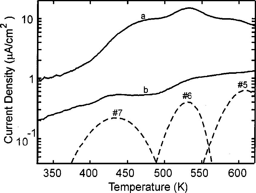
FIG. 2. PICTS spectra measured before (a) and after (b) NI. The calculated spectral lines
corresponding to deep levels Nos. 5-7 are reported in the bottom (dashed lines), with arbitrary
amplitude, for comparison.
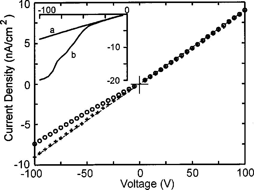
FIG. 3. I – V characteristics before (circles) and after (crosses) NI. The
dashed lines represent the fit of the characteristic after neutron irradiation,
corresponding to a resistivity 1.8×10¹¹ Ω cm. Two curves measured before
NI are shown in the inset. Curve (a) is identical to the curve marked with
circles in the main plot, and is compared with the characteristic measured
after exposure to Xe lamp (curve b).
The reduction of the deep levels concentration determines a general stabilization of the current response. As shown in Fig. 3, before NI, the current–voltage (I – V) characteristic is nonsymmetric with respect to Vbias=0, and shows a linear behavior only for positive biases. This is probably due to polarization effects.12,13 After NI, the I – V curve becomes almost linear and symmetric, denoting a good ohmic behavior, with a resistivity of about 1.8×10¹¹ Ω cm. In the inset, the characteristic measured before NI is compared with the same characteristic obtained after a 30 min exposure to the xenon lamp and a 30 min de-excitation at room temperature. After illumination, the current grows, and the I – V curve becomes highly nonlinear, putting into evidence the occurrence of polarization effects. The initial I – V behavior is recovered after 20 h annealing at room temperature, or after heating the sample at 650 K.
The dosimetric characterization of the sample was carried out at the radiotherapy unit of Dipartimento di Fisiopatologia Clinica of Florence. During exposure, the sample was placed in a cylindrical polymethylmethacrylate housing and biased with Vbias=100 V. The current response of the sample under exposure to a Co60 source (dose rate Dr=0.2 Gy/min) is shown in Fig. 4. To passivate the deeper traps, the sample has been γ-irradiated up to a dose of 20 Gy before measurements. Before NI, the current under γ exposure is steadily increasing during time and, when the beam is switched off, the current shows a slow decay. After NI, the intensity of the on-line current is lowered by one order of magnitude, but the dynamic response is heavily improved. The signal is well reproducible after several irradiation cycles, achieving a stable value within a few seconds. The charge released over each irradiation cycle grows linearly with the absorbed dose, determining a sensitivity of about 45 nC/(Gymm3). This value is definitely higher than the sensitivity of ionization chambers, and compares favorably with those of standard silicon dosimeters and best-quality CVD and natural diamond devices.14,15
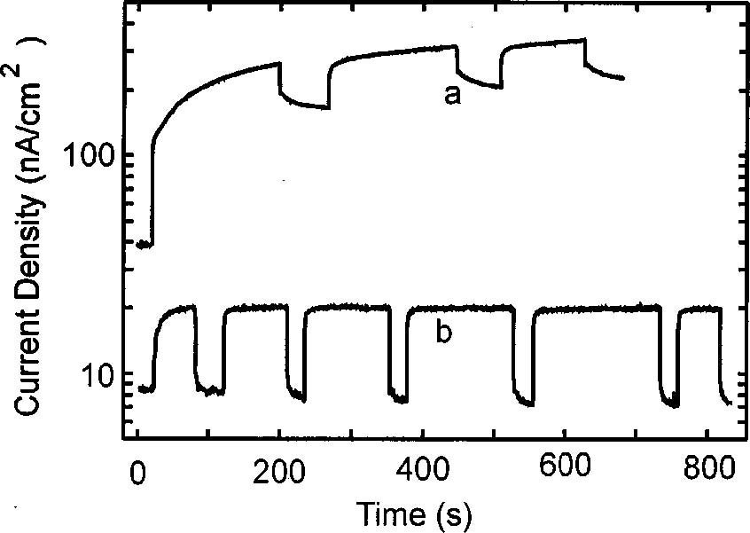
FIG. 4. Current response of the CVD diamond during room temperature exposure to a Co60 γ-source measured (a) before (as grown) and (b) after NI. The source has been switched on and off several times to test the dynamic response of the sample. The applied voltage is 100 V, the dose rate is 0.3 Gy/min.
The current response as a function of the dose rate has been studied using the Co60 source and a 6 MV photon radiotherapy beam from a linear accelerator in the range 0.1–10 Gy/min. Results have been fitted to the standard relationship for on-line dosimeters: I=I0+RDΔr, with Δ=0.9. The quasilinear dependence of the current response with the dose rate suggests that, after NI, a significant residual trap concentration is still present in the material. The microscopic mechanism which causes the radiation-induced removal of the 0.9–1.4 eV levels is presently not clear. Neutron irradiation generates vacancy–interstitial pairs in the material bulk. The capture of mobile self-interstitial at extended defects or grain boundaries has been invoked to explain the decreased intensity of the 2.156 eV luminescence line after neutron irradiation.16 This explanation was based on the assumption that the self-interstitial is mobile at room temperature. However, recent studies17 indicate that the selfinterstitial is not mobile below 700 K. Vacancy is not mobile at room temperature, as well.18 Defects related to vacancy are created by neutron irradiation, giving rise to the well known GR and ND absorption bands.19,20 The shallower vibronic system GR1, which is related to neutral vacancy, is characterized by a zero-phonon line at 1.67 eV; the GR2 to GR8 lines are localized between 2.8 and 3.0 eV, while the ND1 system, associated with the vacancy in negative state, has a zero-phonon line at 3.15 eV. GR2–8 and ND1 transitions are associated with photoconductivity,19 but are not detectable by TSC and PICTS in the temperature range investigated in this work, due to their high activation energy. For the same reason, they should produce a negligible effect on the dynamic response of the device operating at room temperature. Since vacancies and interstitials are not mobile at room temperature, their reaction with existing defects at grain boundaries can not explain the decrease of the TSC and PICTS signals after NI. Nevertheless, the vacancy could act as a deep trap or recombination center for carriers. The deep energy levels created in large concentration by NI could interact with the existing 0.9–1.4 eV levels, significantly reducing their filling under excitation. Moreover, if 0.9–1.4 eV levels are related to extended defects, structural changes subsequent to the direct collision between defects and neutrons might occur.
Our results demonstrate that fast neutron irradiation at high fluence levels can be a very useful tool to optimize the performance of CVD diamond devices, through the controlled tailoring of the deep level distribution in the material bulk. This opens the way for the development of low cost, high-quality CVD diamond on-line dosimeters for clinical applications in radiotherapy.
The authors wish to thank M. Scaringella for technical support. This work has been carried out in the framework of the RD42 (CERN) and of the CANDIDO (INFN) collaborations. The neutron irradiation at ATOMKI was supported in part by the Hungarian Scientific Research Fund (OTKA) (Contract No. T T026184).
1R. J. Keddy, T. L. Nam, and R. C. Burns, Phys. Med. Biol. 32,751 (1987).
2E. A. Burgemeister, Physica B 11, 319 (1981).
3J. Barthe, Nucl. Instrum. Methods Phys. Res. B 184, 158 (2001).
4J. F. Fowler, in Radiation Dosimetry, edited by F. H. Attix and W. C.Roesch (Academic, New York, 1966), Vol. 2, pp. 291–322.
5J. Ruan, K. Kobashi, and W. J. Choyke, Appl. Phys. Lett. 60,3138 (1992).
6D. Takeuchi, H. Watanabe, S. Yamanaka, and H. Okushi, Phys. Rev. B 63, 245328 (2001).
7E. Vittone, C. Manfredotti, F. Fizzotti, A. LoGiudice, P. Polesello, and V. Ralchenko, Diamond Relat. Mater. 8, 1234 (1999).
8D. Tromson, A. Brambilla, P. Bergonzo, A. Mas, C. Hordequin, C. Mer, and F. Foulon, J. Appl. Phys. 90, 1608 (2001).
9P. J. Sellin, M. B. H. Breese, A. P. Knights, L. C. Alves, R. S. Sussmann, and A. J. Whitehead, Appl. Phys. Lett. 77, 913 (2000).
10M. A. Lone, C. B. Bigham, J. S. Fraser, H. R. Schneider, T. K. Alexander,
A. J. Ferguson, and A. B. McDonald, Nucl. Phys. A143,331 (1977).
11 M. Bruzzi, L. Lombardi, D. Menichelli, and S. Sciortino, J. Appl. Phys. 91, 5765 (2002).
12M. Marinelli, E. Milani, A. Paoletti, A. Tucciarone, G. Verona Rinati, M.
Angelone, and M. Pillon, Diamond Relat. Mater. 10, 645 (2001).
13B. Gan, J. Ahn Rusli, Q. Zhang, S. F. Yoon, V. A. Ligatchev, J. Yu, K.
Chew, and Q. F. Huang, J. Appl. Phys. 89,5747 (2001).
14C. De Angelis, S. Onori, M. Pacilio, G. A. P. Cirrone, G. Cuttone, L.
Raffaele, M. Bucciolini, and S. Mazzocchi, Med. Phys. 29,248 (2002).
15C. M. Buttar, R. Airey, J. Conway, G. Hill, S. Ramkumar, G. Scarsbrook,
R. S. Sussmann, S. Walker, and A. Whitehead, Diamond Relat. Mater. 9, 965 (2000).
16L. Allers, A. S. Howard, J. F. Hassard, and A. Mainwood, Diamond Relat.Mater. 6, 353 (1997).
17D. C. Hunt, D. J. Twitchen, M. E. Newton, J. M. Baker, T. R. Anthony, W.
F. Banholzer, and S. S. Vagarali, Phys. Rev. B 61, 3863 (2000).
18G. Davies, S. C. Lawson, A. T. Collins, A. Mainwood, and S. J. Sharp, Phys. Rev. B 46, 13157 (1992).
19J. Walker, Rep. Prog. Phys. 42, 1605 (1979).
20J. E. Lowther, Phys. Rev. B 48, 11592 (1993).
a)Author to whom correspondence should be addressed; electronic mail:
bruzzi@fi.infn.it
0003-6951/2002/81(2)/298/3/$19.00 298 © 2002 American Institute of Physics
Downloaded 05 Aug 2002 to 148.6.160.6. Redistribution subject to AIP license or copyright, see http://ojps.aip.org/aplo/aplcr.jsp