Scanning electron microscope (SEM)
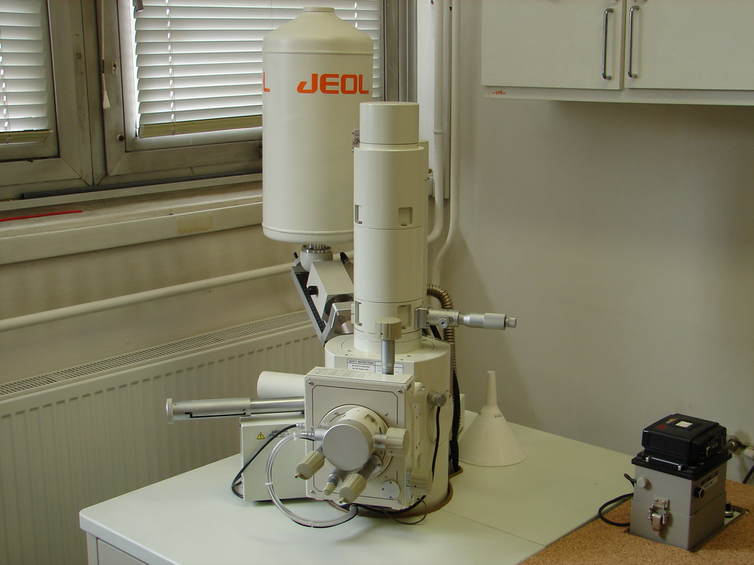
One of the basic instruments of the Radiation Chemistry Department is the scanning electron microscope, JEOL JSM 5600LV. The accelerating voltage of this high-performance microscope can be changed between 0.5 – 30 kV, with magnifications from x35 – 300 000.
This SEM also has a low vacuum operating mode, allowing the observation of specimens that cannot be viewed at high vacuum due to their excessive water content or because of a non-conductive surface. In this low-vacuum mode the samples can be observed without any preparation, thus minimizing the appearance of artifacts.
For high vacuum mode observation nonconductive surfaces can be gold- or palladium/platinum-coated by using the JFC-1300 Auto Fine Coater.
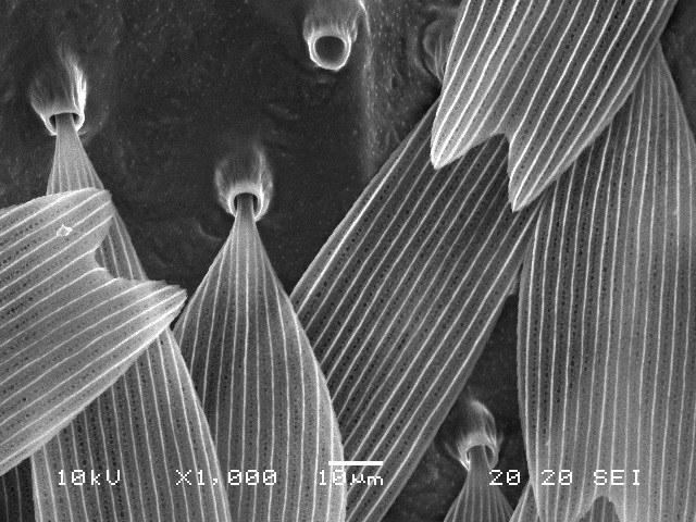
The microscope is regularly used for investigation of polymer morphology: porous structure of hydrogels, sizes of functional micro- and nanospheres, porous structure of polymer monoliths. Besides these, damages introduced by various methods to textile and glass fibers, as well as biological samples are regularly studied.
The microscope is equipped with and EDS2000 Microanalysis system enabling a complete analytical analysis of the sample located in the SEM.